This is a message for foreigners living in or visiting Japan who suffer from homonymous hemianopsia, a condition caused by a stroke!
Good news for those suffering from homonymous hemianopia, which are the after-effects of stroke!
Our clinic specializes in the treatment of homonymous hemianopsia, a sequela of stroke.Ryuei Acupuncture Clinic in Tokyo Japan
At our clinic, we provide effective acupuncture treatment for homonymous hemianopia.
We called Katsu-no-shin【活脳鍼】
If you come to Japan, please come to our clinic. If possible, please bring an interpreter with you.
If an interpreter is not available, you will have to use a translation machine, so detailed explanations of treatment may not be available.
Katsunoshin is an acupuncture treatment that promotes cerebral blood flow, activates the brain, and improves the aftereffects of stroke.
Therefore, it is effective for motor paralysis, sensory paralysis, double vision, homonymous hemianopia, etc. which are by cerebral hemorrhage or cerebral infarction sequelae.
It is particularly effective for refractory homonymous hemianopsia.
Click here for more information about Katsunoshin (Japanese)>>
Homonymous hemianopia is a condition in which the visual field on one side is reduced by half.
The nerves in your eyes optical nerve intersect, so if you lose vision in the left half of your field of vision, there may be a problem with your right occipital lobe. This is called left homonymous hemianopia.
In the case of the right half, the disorder is in the left occipital lobe and is called right homonymous hemianopia.
If you have trouble grasping things or bumping into them, it can seriously interfere with your daily life. I get scared when I can’t see moving cars or bicycles. You can’t even go out to play. However, there is still no effective treatment for homonymous hemianopia caused by cerebral infarction or cerebral hemorrhage.
So, many people suffer from homonymous hemianopia. The reason is that there are no innovative rehabilitation.
Then,we have no choice but to increase the reversibility of the brain.
But, please do not give up!
Our clinic unique acupuncture treatment method, Katsunoushin, promotes blood flow to the brain and activates.
As a result, the plasticity of brain tissue increases, Repair necrotic areas caused by cerebral infarction or hemorrhage.
If you continue using Katsunoushin, it is not uncommon for necrotic areas to disappear.
But, it’s not a miracle.
The effect of activating the brain has been demonstrated by optical topography testing and electroencephalography testing. This is the evidence that Katsunoushin increases brain plasticity.
And also many examples of improvements prove this.
Although it is said that homonymous hemianopsia in particular does not recover, there are many cases in which visual field is restored with Katsunoushin.
If you are suffering from homonymous hemianopia, please try our clinic’s unique Katsunoushin.
Now, I will explain in order the effects of Katsunoushin on the homonymous hemianopia.
What does it mean to watch?
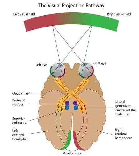
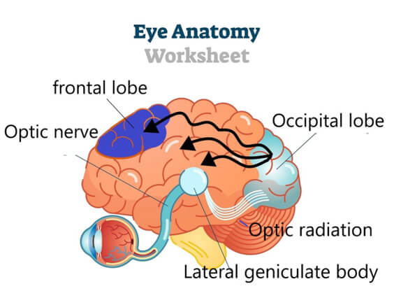
Objects entering the eye form an image on the retina, which is converted into electrical signals and transmitted to the optic nerve.
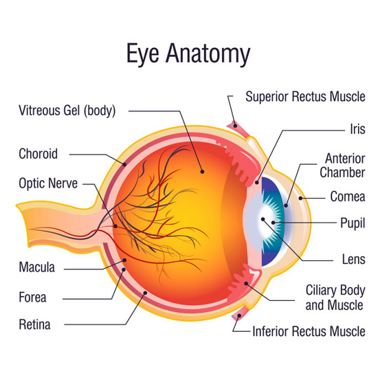
Information crosses at the optic chiasm along the way. When the information crosses, it is recombined from “visual information in the right eye and visual information in the left eye” to “visual information in the right half of both eyes and visual information in the left half of both eyes”.
Furthermore, one is connected to the primary visual cortex of the right occipital lobe and the other to the primary visual cortex of the left occipital lobe, where the information is processed separately, enabling usto recognise what we see with our eyes in 3D.
And, Information is transferred from the primary visual cortex to the higher visual cortex, where image processing proceeds.
The result is an image that accurately describes both shape and colour.
Furthermore, this information reaches the frontal lobe, the thinking brain, via the parietal and temporal lobes.
The frontal lobe determines what the image is and uses it as a reference for action. In addition, the information is stored in the temporal lobe.
When you are watching something while thinking, this is also when your frontal lobe is fully active and trying to make sense of the image.
For example, when we look at an image on a computer screen, we think about it, remember it, and process it appropriately. This is the function of the frontal lobe.
Of course, this requires the occipital lobe to create accurate image data.
This task becomes difficult when the visual cortex in the occipital lobe is damaged by a stroke or brain hemorrhage.
When it spreads widely to the left and right occipital lobes, images cannot be formed at all and visual field is lost.
However, complete blindness is rare, and symptoms such as loss of half of the visual field due to unilateral cerebral infarction or cerebral hemorrhage often occur.
This is called homonymous hemianopia.
What is a field of view?
Field of vision is the area that can be seen when you look at a single point without moving your eyes.
Also, the viewing angle refers to the angle at which you can see without problems when looking diagonally in front of you.
It is said that for one human eye, the horizontal angle is around 100 degrees outward, around 60 degrees inward, around 60 degrees upward, and around 70 degrees downward.
Many causes of narrowing this visual angle are retinopathy, glaucoma, retinitis pigmentosa, and retinal detachment.
These are caused by the eyeball itself, but can also occur due to cerebral infarction or cerebral hemorrhage in the temporal or occipital lobe.
In particular, the occipital lobe creates images, so if this area is damaged by a cerebral infarction or cerebral hemorrhage, images will be missing, resulting in narrowed vision. This is called homonymous hemianopia.
What is homonymous hemianopia?
The image that enters the eyeball reaches the occipital lobe and is converted into a detailed image, but if there is a disorder on the way, it causes visual field disturbances.
It is called homonymous hemianopia and is characterized by loss of visual field on one side.
The name homonymous hemianopia means that the visual field defect is on the same side of both eyes.
Therefore, if the right occipital lobe is damaged, both eyes will lose the left half of their visual field.
Homonymous hemianopia can occur for a variety of reasons.
Causes other than stroke include brain tumors,hydrocephalus, and multiple sclerosis.
There are treatments such as surgery, radiation, and drugs for fatal diseases such as brain tumors, but there is no innovative treatment for stroke.
However, if the condition is caused by the after-effects of a stroke, it is not life-threatening, and is said that it will not get any worse.
Additionally, if brain cells in the right hemisphere, including the occipital lobe, are extensively damaged by a stroke, a symptom called left hemispatial neglect may appear.
You will no longer be conscious of the existence of the left side, so you will frequently hit things on the left side.
Next, I will explain a specific example of homonymous hemianopia. See illustration below.
Homonymous hemianopsia is a disorder of the optic tract from the optic chiasm to the lateral pallidum, the optic radiation to the occipital lobe, and the visual cortex of the occipital lobe.
Disorder ➊ in the image above is an impairment of the optic nerve transmission closer to the eye than the optic chiasm. Visual acuity in the right eye is completely lost.
➋ is a disorder of the optic chord, resulting in loss of vision in the right half of both eyes.
This is the most common form of Homonymous hemianopsia.
➌ is damage to Meyer’s loop, which results in loss of vision in the right upper half of both eyes. The Meyer loop is an optic radiation extending downward from the lateral geniculate body. It dominates the upper half of the visual field.
In the illustration, the disorder is in the left optic chordg or left optic radiation, and right homonymous hemianopia is used as an example, but if it is a right optic chordg or right optic radiation, it will be left homonymous hemianopia.
The above is an explanation of homonymous hemianopia.
It is intractable, but please do not give up.
There is a high possibility of improving homonymous hemianopsia with Katsunoushin acupuncture.
If you feel the limitations of Western medicine, please try Katsunoushin acupuncture. Next, let’s take a look at the mechanism of action of Katsunoushin acupuncture for homonymous hemianopia.
Overview of Katsunoshin
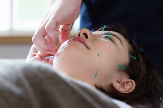
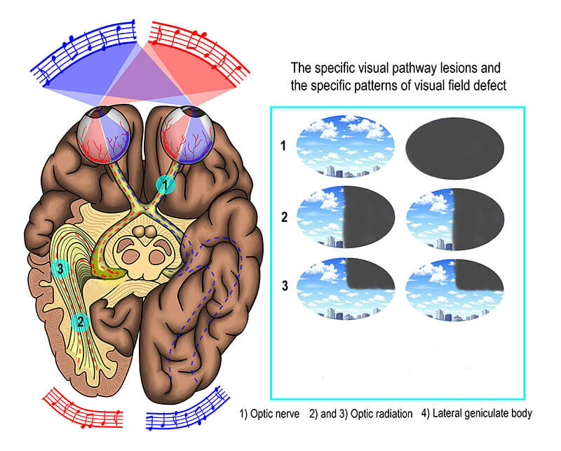
Katsunoshin is a new acupuncture treatment method that combines traditional Chinese acupuncture, Ganshin「眼鍼」, with Ruishin「唇鍼」. It mainly targets the sequelae of stroke. Ganshin inserts acupuncture needles into acupuncture points from the corners of the eyes to the temples. Also, Ruishin involves inserting needles into the skin around the lips. Furthermore, depending on the symptoms, pressure points around the jaw and ears may be used.
Of course, ultra-fine acupuncture needles are used, so you will hardly feel any pain.
However, in rare cases, a thicker needle may be used. In that case, you may feel a dull pain.
But, please be assured that the pain is not unbearable. Well, Ruishin is an acupuncture treatment method that we have developed independently. The reference was the Penfield map.
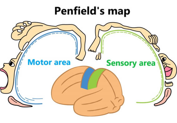
Penfield’s map is a graphic representation of the motor and sensory areas of the brain and their corresponding parts of the body.
As this map shows, the area around the lips occupies a relatively large proportion of the parietal sensory area.
In other words, stimulation around the lips can have a strong effect on the brain.
So, I decided to try using pressure points around the lips as well.
In fact, the addition of Ruisin significantly improved treatment results for homonymous hemianopia.
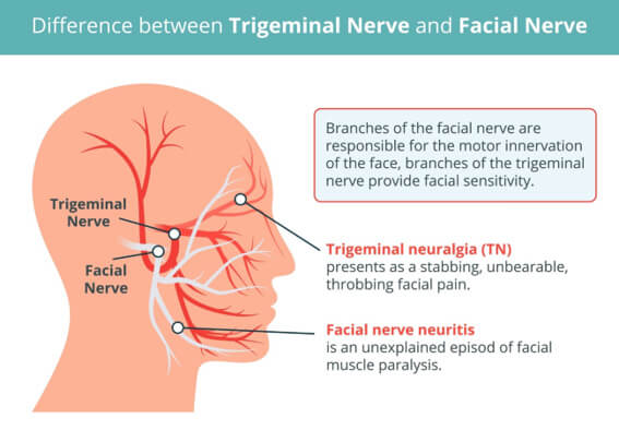
The stimulation from Ganshin is transmitted to the thalamus through the first branch of the trigeminal nerve, which is distributed around the temples and eyes.
Ruishin’s stimulation is transmitted to the thalamus via the 2nd branch of the trigeminal nerve and the 3rd branch of the trigeminal nerve around the lips.
In addition, acupuncture is also applied around the jaw and ears, so that stimulation is also transmitted to the facial nerve and vagus nerve.
Furthermore, the stimulation are transmitted to the parietal, temporal, and occipital lobes via nerves extending from the thalamus.
It also transfers from the thalamus to another nerve and reaches the frontal lobe, including the right dorsolateral prefrontal cortex.
According to our observations, Ganshin seems to awaken the brain, Luisin seems to alleviate it. If the brain is overused, its function is reduced. Relaxation is also necessary. Both interfere with each other and activate the brain. In other words, Katsunoshin has both an excitatory and a sedative effect. This is a very strange phenomenon, since both Ganshin and Luisin stimulate the same trigeminal nerve.
Therefore, I thought that if we could activate the brain, it might also be effective against mental illnesses. When Katsunoshin’s treatment was given once a week for three months without medication, it was found to be effective for depression. Symptoms such as depression, low energy, poor concentration, insomnia, fatigue, and anxiety were greatly reduced.
So, to confirm this, I conducted an electroencephalogram test. The results are as shown in the image below, when I performed Katsunoshin, both alpha and beta waves rose. This suggests that the brain is actively working.
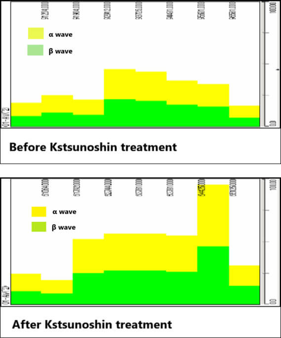
When I looked into the details further, I found that alpha waves are generated throughout the brain when I do luicin, and beta waves are prominent in the frontal and temporal lobes when I do Ganshin.
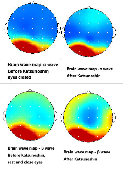
In other words, it can be inferred that Luisin relaxes the mind and Ganshin actively works the brain.
This result cannot be doubted, as similar EEG tests performed on many patients showed the same trend.
Based on these results, we formulated the following hypothesis. Luisin alleviates the discomfort prediction function of the right dorsolateral prefrontal cortex in the frontal lobe, and Gansin activates the left dorsolateral prefrontal cortex and temporal lobe in the frontal lobe, promoting proactive behavior.
It is thought that when humans have a stroke, they try not to move their limbs in order to keep them at rest.
However, this system must be released as soon as possible once the blockage or bleeding in the cerebral blood vessels has subsided.
If it continues for a long time, it can leave behind after-effects such as hemiplegia and speech disorders.
However, once the danger to his life passes and he regains consciousness, he becomes aware that he cannot move his limbs freely.
If this happens, you will develop a “pessimistic belief that your limbs will not move.”
We believe that this is one of the reasons that hinder recovery from hemiplegia.
In other words, the after-effects of stroke are caused by excessive defensive reactions and pessimistic beliefs.
The same goes for homonymous hemianopia. When the occipital lobe is damaged by a stroke, it suppresses its function and attempts to increase its natural healing ability.
However, when he is faced with the reality that he can only see half of his field of vision, he becomes depressed. Furthermore, if a doctor tells you that there is almost no chance of improvement, you will feel a sense of despair.
But, do not give up!
Katsunoshin releases those pessimistic thoughts.
The dorsolateral prefrontal cortex is located on the left and right sides, and the right side is responsible for pessimistic (unpleasant) predictions, and the left side is responsible for positive (pleasant) predictions.
The stimulation of Katsunoshin acupuncture generates alpha waves throughout the cerebrum, including the right dorsolateral prefrontal cortex, which puts the brain in a relaxed state and helps you forget the pessimistic belief that your limbs cannot move.
At the same time, it stimulates the left frontal lobe. It strengthens the beta waves and expresses the intention to “start moving!” This is transmitted to the motor cortex, where the actual action begins.
In addition, hints for movement come from the temporal lobe, which is full of beta waves.
We believe that when the temporal lobe becomes active, it may revive memories of when we were able to move around freely and remind us of how to move our limbs, which we had almost forgotten.
The same logic applies to improving homonymous hemianopia. It puts the right dorsolateral prefrontal cortex into a relaxed state, making you forget the pessimistic belief that you can’t see, and at the same time increases the function of the left frontal lobe, strengthening your intention to ”see!” This is transmitted to the occipital lobe and is the driving force behind restoring visual field. Visual memories are also stored in the temporal lobe. The activated temporal lobe will give you hints on how to expand your field of vision.
Actions and effects of Katsunoshin on homonymous hemianopsia
It is not known exactly how Katsunoshin affects homonymous hemianopia. This is unavoidable, as even the best modern medicine understands only a part of brain functions.
However, at our hospital, we have elucidated the mechanism of action of Katsunoshin acupuncture as much as possible, and based on the information obtained from the results, we have developed a more in-depth hypothesis.
Images entering the eye are transported via the thalamus to the temporal and occipital lobes. They are then recognised by the frontal lobes and clear image processing takes place.
Therefore, functional enhancement of this neurotransmission circuit may increase brain plasticity.
Similarly, the stimulation of Katsunoshin acupuncture is also transmitted to the frontal lobe, parietal lobe, and temporal lobe via the thalamus. Naturally, it is also transmitted to the occipital lobe.
The resultIt is conceivable that the stimulation of Katsunoshin acupuncture may enhance brain plasticity by activating the brain via these neural pathways. This has been found in studies using an instrument called optical topography.
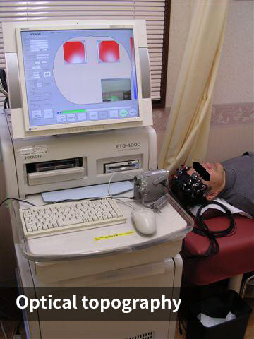
Optical topography is a device that uses infrared light to measure cerebral blood flow.
Blue, white, and red colors indicate cerebral blood flow. Cerebral blood flow is lowest in blue and increases from white to red.
The white image below is before Katsunoshin, and the red image is after Katsunoshin. There is clearly an increase in blood flow.
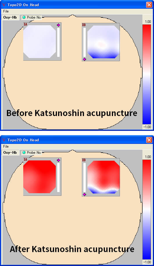
This study was conducted on multiple stroke patients with similar results, so there is no doubt that Katsunoshin increases cerebral blood flow.
It is a common theory that homonymous hemianopsia does not improve, but this is by no means true. Many people experience a widening of the visual field after the first treatment with Katsunoshin acupuncture. In most cases, the visual field is gradually enlarged, to a greater or lesser extent, in the visual field test. Ophthalmologists cannot hide their surprise at this.
For more information, please continue reading.
Detailed explanation of the action and effects of Katsunoshin
Although there are some parts of the content that overlap,we will further explain the effects and effects of Katsunoshin.
Most homonymous hemianopsia is caused by stroke, such as cerebral hemorrhage or stroke, and is due to damage to the occipital and temporal lobes, where the optic chordg and optic radiation run, as described above.
Therefore, to improve symptoms, these neurotransmissions need to be enhanced and the photoreceptor cells activated.

Anatomically, it is assumed that stimuli given to the thalamus are transmitted to the lateral geniculate body, which sends the information to optic radiation of the temporal lobe and then to the primary visual cortex of the occipital lobe.
In addition, there is a pathway from the upper end of the trigeminal principal sensory nucleus of the brainstem to the superior colliculus, which also afferently conveys stimuli to the temporal and occipital lobes.
Based on this nerve transmission, we will consider the effects of Katsunoshin.
When we look at an object, stimulation is transmitted from the thalamus to the occipital lobe, and in the same way, stimulation of the trigeminal nerve is thought to affect the temporal and occipital lobes from the thalamus.This is because this has been confirmed by experiment.
Therefore, Katsunoshin, which strongly stimulates the thalamus, has a large effect on both the temporal and occipital lobes.
Moreover, the stimulation of Katsunoshin is transmitted to the frontal lobe and parietal lobe.
The frontal eye fields and accessory eye fields of the frontal lobe also transmit information directly to the superior colliculus of the brainstem.
In this way, multiple pathways flow visual information to the occipital lobe via the superior colliculus.
On the contrary, the superior colliculus is closely associated with visual impairment. When the frontal lobe stimulates the superior colliculus, there is a greater chance of detecting the presence of an object without going through the occipital lobe.
This means you can avoid obstacles even if you can’t see them.
In other words, the stimulation transmitted from the thalamus to each brain area feeds back and works to stimulate the brainstem. These systems are called the cerebral cortex-basal ganglia loop and the basal ganglia-brainstem system.
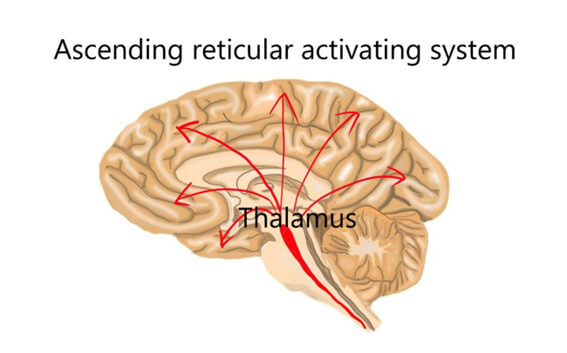
In order to carry these systems smoothly, it is essential to activate the ascending reticular activating system.
Katsunoshin strongly stimulates the thalamus, increasing the possibility of activating the ascending reticular activating system. This can be inferred from the instant improvement in hemiplegia caused by stroke.I also think that .the rise in brain waves that I explained earlier also suggests this.
It goes without saying that stimulating the temporal and occipital lobes is effective in improving homonymous hemianopia, but activation of the frontal lobes is also necessary.
The above is the hypothesis of the mechanism of action of Katsunoshin acupuncture on homonymous hemianopsia.
Here is a case of homonymous hemianopsia, which is more proof than argument.
A visual field test shows that the field of vision is widening.
Clinical results using visual field testing as an indicator of improvement
Regardless of speculation on the mechanism of action of katsunoshin acupuncture for hemianopsia, it is in fact highly effective for motor and sensory paralysis of the limbs, as well as for diplopia and hemianopsia.
Many patients experience a widening of their field of vision even after the first treatment.
There are likewise people who experience an improvement in their diplopia and focus.
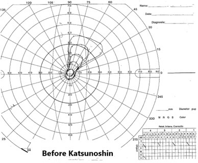
Here is an example of improvement in hemianopsia. The patient had an infarction in the occipital lobe and was homonymous hemianopsia but homonymous quadrantal blindness.
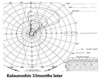
On the contrary, the actual range of vision was even smaller, about 1/10th of that. However, the range of vision has now recovered to about 1/2 of that. The plasticity of the brain, it’s amazing. Please take a look at it.
T.T., 75 years old male
History of present illness
In mid-December 2011, he suffered a myocardial infarction and urgently underwent coronary artery bypass surgery at a university hospital.
A blood clot then flew to his right occipital lobe and he developed left homonymous hemianopia. He first visited our hospital on August 15, 2012. He initially came to the hospital with his family because he didn’t have the confidence to walk alone. He brought the results of the visual field test he had taken on July 9 of the same year. We advised him to have regular visual field tests as an indicator of improvement.
Progress
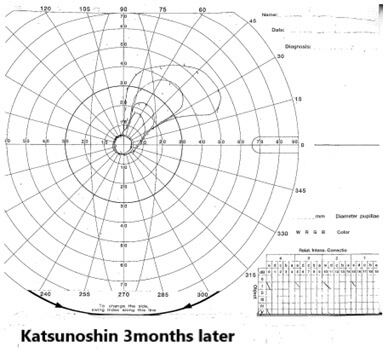
After performing an oriental medical diagnosis, we perform Katsunoshin acupuncture.
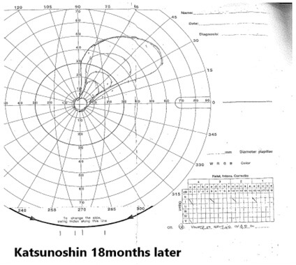
Acupuncture needles were inserted into the area controlled by the first branch of the trigeminal nerve so as to stimulate the occipital lobe in particular. She commented that her surroundings seemed brighter after the treatment. After that, he started having a Katsunoshin acupuncture once a week, and from around the third time onwards, he felt that his field of vision had broadened. Indeed, as shown in the image below, a slight broadening of the visual field was observed during the visual field test on September 5, 2012. After that, periodic visual field tests revealed that his visual field had definitely widened. Lately, I have been hearing happy voices, such as that it is now easier to grab things on the left side, and that they have gained more confidence in walking. Of course, I started coming to the hospital alone. In addition, what is noteworthy about this case is that it improves even at advanced ages, and even after several years have passed.
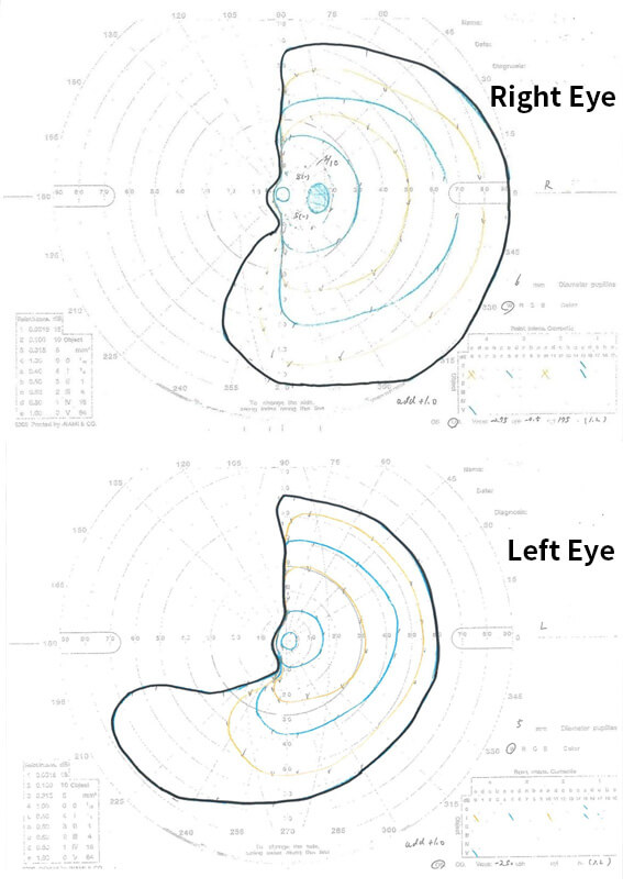
M.S., 40 years old female
History of present illness
In April 2023, he developed left homonymous hemianopia due to subarachnoid hemorrhage. Her doctor told her that it was impossible for her to recover, but she wanted to make some progress, so she and her husband came to the hospital.
progress
Although Katsunoshin’s treatment was carefully performed, she did not notice that her vision had expanded at first and received the treatment with a worried look on her face. However, after 3 or 4 treatments, she felt that his lower left side had widened.
So Ophthalmologist performed a visual field test on her and found that her symptoms were clearly improving. In the image below, the solid blue line is her visual field at the time of onset, the yellow line is her visual field 2 months after the acupuncture treatment, and the black line is her visual field at 5 months. Within about six months, the visual field in both eyes had expanded.
The recovery of her visual field below her left eye was particularly remarkable. There are cases in which conditions that are considered unrecoverable by Western medicine can be improved by Oriental medicine. never give up.
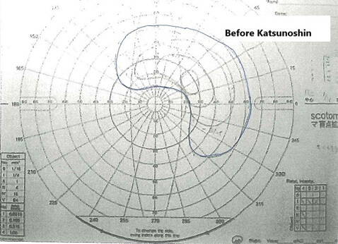
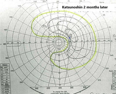
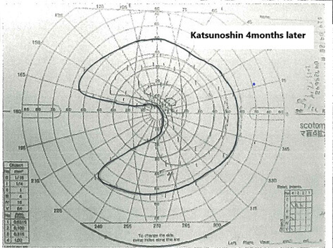
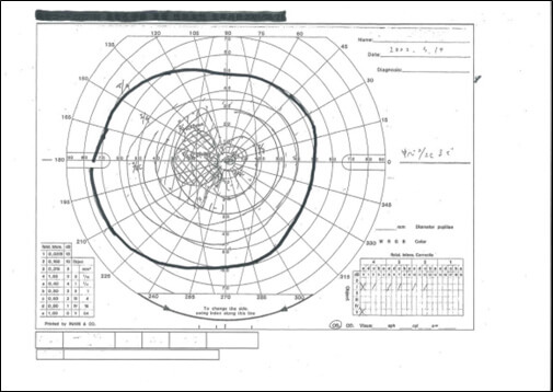
S.Y., 83 years old male
History of present illness
The polyp compressed the optic nerve in the orbit, resulting in unilateral blindness. He had surgery to remove it in February 2023 and was on the mend, but even after one month, he still had lost nearly half of his visual field (blue line in the figure below). In particular, the lower part was severely damaged. So, on April 7th, he traveled a long way to visit the hospital.
progress
In addition to Katsunoshin acupuncture, he also performed acupuncture treatment for glaucoma. From the first visit, he was happy that his horizons had expanded. In fact, a visual field test 2 months after treatment revealed a significant expansion of the visual field (yellow line in the image below).
By the fourth month, his defect had become so small that he felt no discomfort at all (black line in the figure below). Currently, he continues to visit the hospital in hopes of making a full recovery.
Based on these results, we feel that it is necessary to promote fundus blood flow and improve optic nerve function when the optic nerve is injured from the optic chiasm to the eyeball.
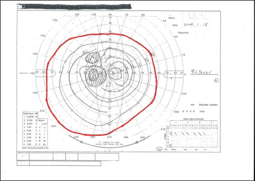
K.S., 20years old female
History of present illness
One year ago, she suffered a cerebral hemorrhage in the left occipital lobe, resulting in right homonymous hemianopia. Her doctors told her there was “no chance of her recovery” and she became depressed.
So, hoping to improve her symptoms even a little, she decided to undergo Katsunoshin treatment about twice a month.
A few months later, we encouraged her to have her vision tested at her hospital because she felt like her vision was improving.
Test results in January 2024 showed that her lower visual field had clearly expanded, as shown by the red line. It is obvious when you compare it with the black line in May 2022.
Above, we have explained the effects of Katsunoshin on visual field disorders. To date, we have treated hundreds of cases, most of which have improved.
It is a well-established theory that homonymous hemianopia does not improve in Japan or overseas, but this is incorrect.
The reason for this is that Western medicine is not good at treating homonymous hemianopia. If you receive Katsunoshin treatment, there is a high possibility that your field of vision will expand.
Profile
| 商号 name | りゅうえい鍼灸治療院 Ryuei Acupuncture Clinic |
| 所在地 address | 東京都三鷹市上連雀2丁目3番12号 2-3-12 Kamirenjaku, Mitaka-shi, Tokyo  |
| アクセス Access | JR中央線三鷹駅南口徒歩5分 4minute walk from the south exit of Mitaka Station on the JR Chuo Line |
| 電話 Phone number | TEL: 0422-40-0077 Japan 0422-40-0077 |
| 創業 Date of establishment | 1989年8月18日 Founded: August 18, 1989 |